Bridging Spatial Biology & Research Excellence in South Africa
Partnering BIOCOM Africa and Miltenyi Biotec to Unlock 3D Tissue Structure and High-Plex Molecular Insight
In modern life-science research, answering complex biological questions increasingly demands both spatial context and deep molecular profiling. The collaboration between BIOCOM Africa (the local distributor supporting labs across southern Africa) and Miltenyi Biotec (a global provider of advanced spatial biology instrumentation) offers a compelling pathway for labs to elevate their workflows. Let’s explore how two of Miltenyi’s key platforms — the UltraMicroscope Blaze™ and the MACSima™ Platform — can transform your research, and how BIOCOM Africa enables access, support and local service in South Africa.

1. Who is BIOCOM Africa & Why This Partnership Matters
BIOCOM Africa positions itself as a provider of cutting-edge laboratory products, offering broad instrument, reagent and consumable portfolios for research labs in southern Africa. Crucially, BIOCOM Africa is listed as the South African distributor for Miltenyi Biotec.
That means for researchers in South Africa:
*Local access to advanced spatial biology instrumentation (reducing import/logistics complexity).
*Local technical support and service infrastructure (important for uptime and throughput).
*Ability to integrate global technology (Miltenyi’s platforms) with local research priorities (disease models relevant in SA, translational pipelines).
So, when you pair BIOCOM Africa + Miltenyi Biotec, you get not just the tools, but the support ecosystem to make them effective in your lab environment.
2. Miltenyi Biotec’s Spatial Biology Portfolio: A Snapshot
Miltenyi Biotec offers a comprehensive “Imaging & Spatial Biology” portfolio: from sample staining, imaging instrumentation, analysis software and high-plex antibodies.
Two flagship platforms in this portfolio are:
The UltraMicroscope Blaze™ (a 3D large volume / light sheet imaging system)
The MACSima™ Platform (a fully automated cyclic immunofluorescence system for ultra-high-plex spatial proteomics)
Together, these platforms enable workflows that span anatomy → molecular phenotypes → spatial context.


")

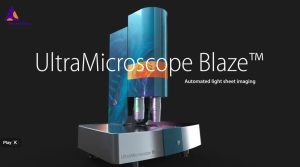
3. UltraMicroscope Blaze™ – Exploring 3D Tissue Architecture
What it does
Uses light-sheet microscopy to image large cleared samples (whole organs, thick tissues) in 3D with subcellular resolution.
Supports high throughput: the UltraMount sample carriers, optimized optics and software streamline large volume imaging.
Provides the anatomical/structural context: you can visualise vasculature, cell networks, tissue architecture in 3D.
How this improves your research
Enables you to move beyond 2D slices: seeing the full context of cell populations in intact tissue helps avoid sampling bias.
Enhances decisions about ROIs: you can decide where to interrogate molecules based on a 3D map.
Supports studies in complex tissues (brain, tumour, organoids) where spatial relationships are key.
Enables richer publications: visually compelling 3D renderings + quantitative volumetrics.
Key considerations
Tissue clearing and sample preparation become critical: large volume imaging demands consistent clearing, fluorescence preservation, refractive index matching.
Data size & storage: 3D imaging generates large datasets — plan infrastructure.
Integration downstream: once you’ve imaged in 3D, you need workflows (sectioning or downstream analysis) to link to molecular data.


")
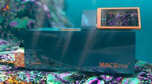
4. MACSima™ Platform – Ultra-High-Plex Spatial Biology
What it does
The MACSima Platform supports same-section multiomics: hundreds of protein markers (and optionally RNA probes) on a single sample.
Built around cyclic immunofluorescence — staining, image, remove cycles — fully automated.
Advanced analysis software (MACS® iQ View) supports segmentation, clustering, gating and multi-marker visualization.
How it improves your research
Deep phenotyping: you can interrogate complex cell types, states, signaling phenotypes in situ.
Spatial context: unlike dissociated single-cell methods, you retain tissue architecture and interactions.
Streamlined workflow: automation reduces labour, increases reproducibility, and aids scaling.
Ideal for translational science: tumour microenvironment (TME), immune cell niches, developmental biology, stem cell niches.
Key considerations
Panel design & QC: ultra-high-plex means you must carefully design antibody cycles, verify removal/re-staining efficiency.
Data management & analysis: high-plex imaging = high-dimensional data; require analysis pipelines and expertise.
Sample throughput: although automated, cycle times and instrument queueing need planning if many samples.
5. The Synergy: Blaze + MACSima = Structural + Molecular Depth
Workflow integration
Use UltraMicroscope Blaze to scan your cleared tissue → obtain a 3D map of structure, identify regions of interest (ROIs).
From the 3D map, select target regions (e.g., tumour-invasion front, vascular niche, stem cell niche).
Section those ROIs (or extract) and apply MACSima to perform high-plex staining/imaging, obtaining detailed molecular maps in the chosen region.
Integrate: overlay structural 3D context with high-plex molecular data → deeper biological insight.
Why this matters
The major gap in many labs is the link between macro-structure and micro-molecular state. Using both platforms bridges that gap.
Greater confidence in ROI selection: rather than choosing arbitrary slices, you’re guided by 3D maps.
Enhanced storytelling: when publishing or presenting, you can show how the macro structure (via Blaze) and micro phenotypes (via MACSima) correspond.
Improved translational value: e.g., in tumour research you can map the architecture, then profile the cells at specific invasive fronts or immune niches with high multiplexing.


6. What This Means for Researchers in South Africa / Africa
Thanks to the BIOCOM Africa–Miltenyi partnership:
Local access: labs in South Africa (and southern Africa) can engage with these advanced tools without full import complexity.
Local support & training: Miltenyi’s training courses (via Miltenyi University) include sessions on the UltraMicroscope Blaze and MACSima workflows.
Relevance to regional research: whether you’re investigating infectious disease, cancer, developmental models or organoids relevant to Africa, these tools scale.
Future-proofing your lab: investing in spatial biology platforms positions your lab for collaborations, funding and publications in a global research environment.
7. Getting Started: Practical Steps
Step 1: Define your research question
Do you need 3D architecture (whole organs/large tissues) or high-plex molecular maps (cell phenotyping)? Or both?
Clarify sample type, model, output expectation (publication, translational pipeline, target discovery).
Step 2: Engage with BIOCOM Africa / Miltenyi for demo & workflow consultation
Discuss sample types, throughput, budget, support.
Arrange training (Miltenyi University offers hands-on & online) for Blaze & MACSima.
Step 3: Pilot your workflow
For Blaze: clear a sample, image at low resolution, test mounting and imaging.
For MACSima: design a small panel, run a pilot cyclic staining, test analysis software.
Step 4: Scale up and integrate workflow
Once pilot is successful: ramp up sample numbers, build data pipelines, plan storage/analysis.
For combined workflows: overlay 3D-to-section workflow, build protocols for ROI transfer.
Step 5: Leverage your data
Produce richer figures for grants/publications: 3D anatomy + high-plex maps.
Use spatial/molecular insight to identify biomarkers, cell niches, therapeutic targets.
Present your lab as spatial-biology capable — enhancing collaboration opportunities.
8. Conclusion
The collaboration between BIOCOM Africa and Miltenyi Biotec empowers South African labs to step into the frontier of spatial biology — not just in isolated techniques, but in integrated workflows that combine structure and molecular detail. By adopting the UltraMicroscope Blaze and MACSima Platform, you can shift from what cells are present to where they are in 3D and how they interact at a molecular level.
For researchers aiming to push the boundaries of tissue-based science — be it tumour microenvironment, developmental biology, infectious disease pathology or organoid research — this pathway offers a game-changing opportunity. With local support, global technology and spatial-biology workflow readiness, the only limit is your scientific question.
Contact
Get In Touch
Every laboratory is unique, and so are its needs. Our team of experts is dedicated to understanding your specific requirements and providing tailored solutions.

